PennHIP Screening in Houston and Spring, TX
At Best Care Animal Hospital in Houston, TX, we specialize in PennHIP which is a Pennsylvania Hip Improvement program at the University of Pennsylvania. PennHIP is a multifaceted radiographic screening method for hip evaluation.
- Si necesita vacunas o un programa económico de Medicina preventiva llame al Hospital Best Care or revise nuestro (PMP) Programa de Medicina preventiva, con precios muy favorables y cuotas desde $20.99 al mes.
- Contamos con programas de pagos que incluyen: servicios Medicos y de laboratorio, Dentisteria, control y tratamiento de parásitos Internos y Externos. Radiología digital, ultrasonido y exámen Avanzado para la detección de la displasia de cadera (PennHIP) en pacientes desde las 16 semanas de edad.
- Hable con el doctor y/o Enfermeras acerca de La Salud de su Mascota en Españo.
Learn More About PennHIP
THE PROBLEM
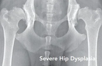
Canine Hip Dysplasia (CHD)
- Is the most commonly inherited orthopedic disease
- Leads to hip arthritis causing pain, stiffness, and diminished quality of life
- Has no medical or surgical cure
- Afflicts more than 50% of the dogs within some breeds
- Clinically affects large breed dogs more severely than smaller breed dogs

THE KEY FACTOR
Hip Laxity
In the 1980’s, researchers at the University of
Pennsylvania’s School of Veterinary Medicine
pioneered a better diagnostic method to assess
hip laxity—the key factor in the development of
Canine Hip Dysplasia (CHD).
The hip joint is a ball-and-socket joint, with the ball of the femur (femoral head) fitting into the hip socket (acetabulum). Hip laxity refers to the degree of “looseness” of the ball in the hip socket.
Studies have shown that dogs with looser hips (excessive hip laxity) are at higher risk to develop hip dysplasia than dogs with tighter hips (minimal hip laxity).
THE SOLUTION
AIS PennHIP Hip Improvement Program
The research-based hip-screening procedure
known as PennHIP has proven to be the most
accurate and precise method to measure hip laxity.
It can identify
—as early as 16 weeks of age—dogs
that are susceptible to developing hip dysplasia.
This offers breeders the opportunity to make
early decisions on breeding stock, and allows veterinarians to advise pet owners on lifestyle
adjustments and preventive strategies to minimize
the pain and progression of the disease.
THE PennHIP PROCEDURE
There are two principal innovations in the PennHIP method. First, the dog is positioned on the x-ray table with hips in a neutral orientation, and second, a custom distraction device is applied to reveal the maximum amount of hip laxity. To achieve this, the dog’s muscles are completely relaxed by administering sedation or general anesthesia.
Veterinarians must complete specialized training and quality-control exercises before becoming certified to perform the PennHIP procedure.
A complete PennHIP evaluation includes office consultation, sedation/anesthesia, and submission of the three PennHIP radiographs to ANTECH Imaging Services for evaluation.
Your certified PennHIP veterinarian will be happy to discuss the procedure and cost with you.

PennHIP RADIOGRAPHS
PennHIP screening includes three separate radiographs (x-rays). Above are examples of the three PennHIP radiographs of a 15-month-old Labrador Retriever.
HIP-EXTENDED RADIOGRAPH(1)
The dog’s hind legs are placed in “extension.” PennHIP utilizes the hip-extended view to identify radiographic signs of hip arthritis also known as osteoarthritis (OA).
Traditional hip screening methods rely solely on the hip-extended view (photo 1) to evaluate both the presence of hip arthritis and joint laxity (subluxation). Using traditional systems this dog’s hips would be considered normal because the hip-extended view (photo 1) shows no evidence of arthritis or subluxation (laxity). While the hip-extended view can detect existing arthritic changes, it often conceals hip laxity thereby giving a false impression of joint tightness. So, in the absence of arthritic changes, as in this dog, the hip-extended view does not reliably distinguish between dogs that are diseasesusceptible and those that are not.
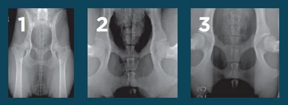
COMPRESSION RADIOGRAPH(2)
The dog’s hind legs are positioned in a neutral, weightbearing orientation and the femoral heads (balls of the femur) are gently seated into the acetabula (hip sockets).
This view can identify critical anatomic landmarks of the hip and determine how well the femoral head fits into the acetabulum.
DISTRACTION RADIOGRAPH(3)
The dog’s hind legs are positioned in the same neutral position as the compression radiograph and a special device is used to reveal the dog’s inherent joint laxity.
This exclusive feature of the PennHIP procedure permits accurate measurement of maximal hip laxity.
When comparing this dog’s hip-extended view (photo 1) to the distraction view (photo 3), the distraction view reveals much greater joint laxity. The PennHIP method uses the amount of joint laxity revealed in the distraction view (photo 3) to tell us that this dog is actually susceptible to developing hip dysplasia and will likely show radiographic evidence of hip arthritis later in life.
HIP SCORING AND REPORT
INTERPRETATION
Your PennHIP veterinarian will submit the three PennHIP radiographs to ANTECH Imaging Services for specialized evaluation. A confidential report comprised of the following key parts will be sent to your PennHIP veterinarian:
Distraction Index (DI)
The DI is a measure of hip laxity—the inherent
distance the ball can be displaced (distracted)
from the hip socket—and is expressed as a number
between zero and one. A DI near zero indicates
little joint laxity (very tight hips). A DI closer to 1.0
indicates a high degree of laxity (very loose hips).
Dogs with tighter hips are less likely to develop
hip dysplasia than dogs with looser hips. A
threshold level of 0.30 has been identified, below
which hip dysplasia is very unlikely to occur.
Arthritis
The PennHIP report also includes an evaluation
of the hip-extended radiograph for evidence of
arthritis, confirming a diagnosis of hip dysplasia.
For dogs with evidence of arthritis, your PennHIP
veterinarian can explain the disease fully and
recommend palliative measures.
Breed Laxity Profile Ranking
Based on the DI, your dog is ranked within its
breed. For the dog breeder this ranking helps in
the selection of breeding candidates—dogs in the
tighter half of the population are recommended
for breeding.
By selecting breeding dogs with tight hips (lower DI), meaningful progress toward better hips can be made within a few generations.
PennHIP — MAKING A
DIFFERENCE
PennHIP is the most accurate hip screening method available and can be safely performed on dogs as young as 16 weeks of age. An early estimate of a dog’s hip integrity is invaluable, whether the dog’s intended purpose is for breeding, for working, or as a family pet.
For breeders
Information compiled in PennHIP’s international
database permits informed selection of breeding
stock based on hip tightness relative to other
members of the same breed. Breeders can
reduce the incidence and severity of Canine Hip
Dysplasia (CHD) in future generations of dogs by
applying selection pressure towards tighter hips.
Among current hip screening methods, PennHIP
has the highest heritability value to bring about
these genetic changes.
For service and working dog organizations
Service and working dog organizations were the
first to adopt PennHIP as the principal method
for hip screening. The investment in training
service/working dogs is enormous. The ability
to prescreen the dog’s genetic predisposition
to CHD is an invaluable tool when evaluating a
future service/working dog’s hip integrity.
For companion dog owners
If your dog is identified to be at risk for CHD,
your PennHIP veterinarian can recommend,
at an early age, appropriate strategies (diet,
medication, and/or activities) to delay or diminish
the ultimate course of the disease.